
Neural system of the mammary gland
Milk ejection is important during milking or suckling to obtain the alveolar milk fraction, which can represent more than 80% of the milk stored in the udder of dairy cows. In response to tactile teat stimulation, either manually or by the milking machine, milk ejection is induced by the release of oxytocin, release from the pituitary gland, resulting myoepithelial contraction. The time from the start of a tactile stimulation until the occurrence of milk ejection spans between 40 s to >2 min and increases with decreasing degree of udder filling. Milk ejection is disturbed under several conditions such as during milking in unfamiliar surroundings or for several weeks immediately after parturition in primiparous cows. Disturbed milk ejection is due to a reduction of or absence of oxytocin.
The milk ejection reflex (let-down) actually is a neuroendocrine reflex (Figure 3). The reflex has an afferent pathway (neural) and an efferent pathway (hormonal, blood-borne). Few nerves go to the interior of the udder. That means that performing a biopsy of the gland to collect tissue can be done with only local anesthetic administered to the skin.
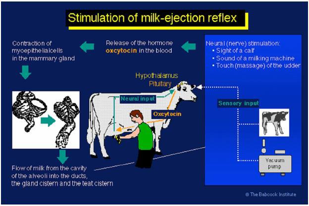
Afferent Pathway
The greatest amount of innervation in the mammary gland is in the teats, where there are pressure sensitive receptors in the dermis. Mechanical stimulation of the teats activates pressure sensitive receptors in the dermis where the pressure is transformed into nerve impulses that travel via the spinothalamic nerve tract to the brain. These nerves synapse in the paraventricular nucleus and in the supraoptic nucleus in the hypothalamus. When the cell bodies of the oxytocin-containing neurons are stimulated by these impulses originating in the teat, an action potential moves down the oxytocin-containing neurons from the cell body in the hypothalamus down the axon to the neuron ending in the posterior pituitary. This causes release of oxytocin into the blood. The efferent pathway starts at this point.
Efferent pathway
The efferent pathway begins with the release of oxytocin into the blood (Figure 3). The oxytocin then travels to the mammary gland via the blood, binds to oxytocin receptors on the myoepithelial cells, causing the myoepithelial cells to contract, and resulting in increased intra-lumenal (intramammary) pressure and ejection of milk from the alveolar lumen. Oxytocin receptors are associated with the myoepithelial cells, not the smooth muscle of the mammary gland.
Other mechanisms of milk ejection:
Myoepithelial cells will also contract in response to vasopressin (ADH or antidiuretic hormone). Vasopressin has about 20% the oxytocic activity of oxytocin;
Visual or auditory stimuli can cause milk ejection. Milk ejection is a condition response;
Stimulation of the genital tract such as vaginal distention causes release of large amounts of oxytocin;
The mechanical tap stimulus does not involve oxytocin. It will occur under anesthesia or denervation of the udder. It is not inhibited by epinephrine. Kneading or butting of the udder by the young may elicit this response. This may involve distortion of the alveolar structure or the myoepithelial cell structure, resulting in milk ejection.
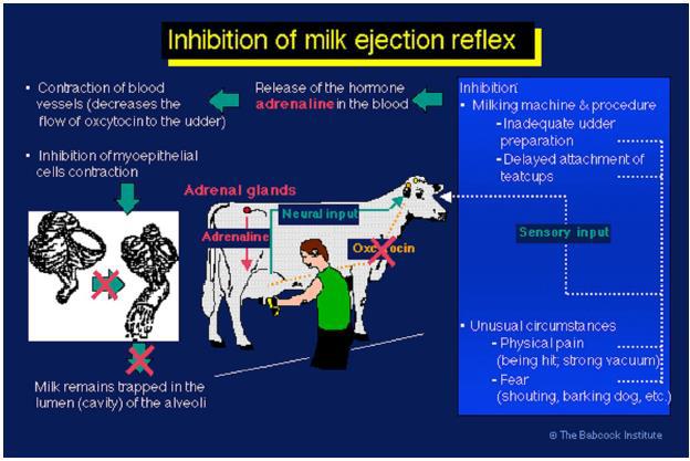
Inhibition of milk ejection
Various stressful stimuli that inhibit milk ejection are associated with increased activity of the sympathetic nervous system. Oxytocin action can be blocked by the hormones catecholamines, which are made by the adrenal glands localized above the kidneys. The main catecholamines are dopamine, norepinephrine and epinephrine (which used to be called adrenalin). These hormones are released in response to stressful situations and increase the tone of the smooth muscles of the mammary ducts and blood vessels. This results in the reduction of oxytocin reaching the myoepithelial cells and partial occlusion of the mammary ducts (Figure 4). Moreover, epinepherin directly blocks oxytocin from binding to myoepithelial cells. This is termed peripheral inhibition of milk ejection. Therefore, in animals exhibiting peripheral inhibition a dose of exogenous oxytocin will not cause milk ejection
A common cause of failure to milk ejection is associated with stress of milking in the early postpartum period especially for primiparous cows. The stress inhibits the release of oxytocin from the posterior pituitary gland. This is termed central inhibition of milk ejection. Exogenous oxytocin is usually administered in these cases causing milk ejection. Based on the above discussion about peripheral and central inhibition of milk ejection, it can be stated that milk ejection occurs as a result of oxytocin release, which is normally couples with inhibition of the central and peripheral inhibitory controls.


