
Reproductive male tracts: anatomy and function
The primary organ of reproduction in the male is the testicles, of which the bull has two, as is the case in most animals. Secondary sex organs and three accessory sex glands are part of reproductive male tracts. All reproductive organs work in concert for formation, maturation and transport of spermatozoa, which are eventually deposited in the female reproductive tract. The secondary sex organs are the epididymis, vas deferens and penis. The three accessory sex glands include the seminal vesicles, prostate and bulbourethral gland (Cowper's gland). The reproductive male anatomy is showed in Figure 3. | 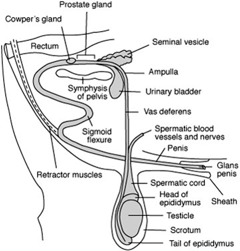 |

The testicles
The testicles are located outside the body cavity in the scrotum (external oval sac) and have two vital functions: producing the spermatozoa, and producing the testosterone (male hormone). Location of the testicles exterior to the body cavity is essential for normal sperm formation, which occurs only at 1 to 4°C cooler than body temperature. The scrotum provides physical protection to the testicle and helps regulate the temperature for optimum spermatozoa development. This regulation is done by coordination of three structures: a temperature-sensitive layer of muscle (tunica dartos) located in the walls of the scrotum, which relaxes when hot and contracts when cold; the external cremaster muscle within the spermatic cord, which controls the proximity of the testicle to the body by lengthening or shortening depending on environmental temperature; and a counter-current temperature exchange regulated by a blood flow process known as the pampiniform plexus, which is a coil of testicular veins that provide an effective mechanism for cooling arterial blood entering the testicle and transferring its heat to the venous blood leaving the testicle.
One or both testicles occasionally fail to descend into the scrotum during embryological development and are retained in the body cavity. This condition is known as chryptorchidism. Hormone production by chryptorchid males is near normal and the male develops and behaves like a normal male, however will generally be subfertile. This condition is genetically inherited, therefore such males should not be used for breeding.
The seminiferous tubules are lined with germinal epithelium, the spermatogonia. The tubes seminiferous are formed by many long, tiny, coiled tubes, within which the sperm are produced and begin to mature. Specialized cells, known as Sertori cells, localized in the tubes seminiferous are responsible to provide nutriments to the spermatozoa. Scattered throughout the loose connective tissue surrounding the seminiferous tubules are other kind of specialized cells, the interstitial cells of Leydig, that produce testosterone. There are hundreds of individual seminiferous tubules in the body of the testicle which unite with one another to form a few dozen tubules that exit from the testicle and pass into the epididymis.
The epididymis
The epididymis is a compact, flat, elongated structure closely attached to one side of the testicle. It is divided into three regions, the head, body and tail. The many tubules entered the head of the epididymis from the testicle unite to form a single tubule. An epididymis bull has approximately 35 meters long and this tubule is convoluted and packed into 18 cm. Four major functions occur in the epididymis, including the transport of the developing sperm cells from the testicle to the vas deferens; the concentration of the sperm by absorption of surplus fluids; the maturation of the developing spermatozoa; and the storage of viable sperm cells in the epididymis tail. If sexual activity is slowed, resorption of sperm cells from the epididymis tail occurs.
The epididymis serves as an outlet for all the sperm produced in the testicle and any blockage of this tube will cause sterility. Temporary blockage due to swelling following an injury or infection (epididymitis) will result in short-term infertility. If the swelling or infection results in formation of scar tissue in the tubule, it may permanently block the passage of sperm. If blockage occurs in both epididymides, the bull will no longer be useful as a breeder. Surgical removal of the tail of the epididymis (epididectomy) is frequently used as a means of sterilization for teaser (Gomer) bulls for estrus detection. Epididectomized bulls will mate cows in the usual manner, because they still produce testosterone from cells Leydig, however will not deposit sperm in the female reproductive tract.
The vas deferens
The vas deferens, also known as ductus deferens, emerges from the tail of the epididymis as a straight tubule and passes as part of the spermatic cord through the inguinal ring into the body cavity. Spermatozoa are transported further along the reproductive tract to the pelvic region through the vas deferens by contraction of the smooth muscle tissue surrounding this tubule during ejaculation. Bulls may also be sterilized by a vasectomy in which a section of the vas deferens is removed so that sperm cannot pass to urethra and go to outside of the body.
The urethra is a single tube that communicates with two vas deferens. Anatomically the urethra is the channel passing through the penis. The urethra serves as a common passageway for semen from the reproductive tract and urine from the urinary tract.
As mentioned above, the bull has three sets of accessory glands. Semen is made up of the fluids from accessory glands and the sperm (mature spermatozoa). The volume of ejaculate is very variable, 1 to 15 ml. The concentration will also vary considerably, however the usual is 1 to 1.8 billion sperm per ml. Since only one sperm is required to fertilize an ovum, considerable dilution of the semen can be done.
Two of accessory glands, the seminal vesicles and prostate, are located in the region where the vas deferens unite to become the urethra. Secretions from these glands make up most of the liquid portion of the semen. In addition, the secretions activate the sperm to become motile. The seminal vesicles consist of two lobes about 10 cm long in the bull, each connected to the urethra by a duct, which the main role is the production of nutrients for spermatozoa. The prostate gland is located at the neck of the urinary bladder where it empties into the urethra. The prostate is relatively small in the bull, as compared to other species, and does not produce a very large volume secretion, which is rich in enzymes for spermatozoa metabolism.
The third accessory gland, the Cowper's glands, are small, firm glands located on either side of the urethra. The clear and buffered secretion that often drips from the penis during sexual excitement prior to service is largely produced by these glands and serves to flush and cleanse the urethra of any urine residue that may be harmful to spermatozoa. The secretion from Cowper's glands assures an optimizer pH for the semen. This is a protection against an eventual low pH of female reproductive tracts and pH decrease due the spermatozoa metabolism.
One of the accessory glands may occasionally become infected, resulting in semen samples that are yellow and cloudy and which contain puss cells. It is not uncommon in bulls for the seminal vesicles to be so affected (seminal vesiculitis). Antibiotic treatment is sometimes necessary, but time will generally correct the problem.
The penis
The penis of bulls has sigmoid flexure. This is an anatomical structure that provides a means by which the penis is held inside the sheath except during time of mate. Strong retractor muscles hold the penis in the "S" shaped configuration. Occasionally these muscles are too weak to function properly and a portion of the penis and sheath lining protrude at all times. This exposes the male to the danger of injury and this characteristic should be avoided when selecting a herd bull.
The penis is the organ of insemination. Spongy-type material within the penis is filled with blood during sexual arousal, resulting in erection of the organ. The end of the penis is the glans penis and is richly supplied nerves, which are stimulated during copulation to induce ejaculation.
The normal functions of male reproduction are largely controlled by hormones that are secreted from the endocrine glands. The testicle functions as an endocrine gland because of its production of the male hormone, testosterone, by the interstitial cells (cells Leydig).
The major functions of testosterone are:
It is largely responsible for development and maintenance of the male reproductive tract;
It causes the development and maintenance of the secondary sex characteristics associated with masculinity, such as the crest and heavily muscled shoulders of a bull;
It is a major factor in the normal sex drive and behavior of the male;
It increases muscular and skeletal growth, and
It is essential for normal sperm formation.
The same gonadotropic hormones that regulate ovarian functions in the cow also regulate testicular functions in the bull. LH and FSH are released from the pituitary gland and cause the testicle to secrete testosterone, which then acts on the germ cell lining of the seminiferous tubules to stimulate formation of primordial sperm cells. The maturation of spermatids into fully developed sperm cells requires the presence of FSH. Normal functioning of the male accessory glands requires testosterone.
Not only is hormone production of the testicle regulated by hormones released by the anterior pituitary, but the reverse is also true. The level of testosterone in the blood regulates the secretion of gonadotropic hormones from the anterior pituitary via a feedback system. A proper balance of all hormones is vital to successful reproductive functions.


