
Reproductive female tracts: anatomy and function
Two essential organs of reproduction are located within the head of the animal.
The hypothalamus controls several body processes and behaviors along with reproductive processes. Body temperature, concentration and components of body fluids and the drive to eat and drink are just a few functions of the hypothalamus. It is classified as a neuroendocrine gland since it sends and receives neural signals through the nervous system and hormonal messages through the endocrine system.
The second organ, the pituitary small gland located at the base of the brain. The pituitary is about half an inch in diameter and weighs about 1 gram. Physiologically, the pituitary is divided into two distinct regions: the anterior and posterior pituitaries. Each region secretes various hormones that direct body processes. Some of these hormones are responsible for reproductive events, while others control growth, metabolism and water balance.
We can distinguish three fundamental functions of female reproductive tract :
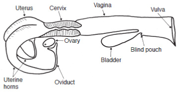
These female steroid hormones are involved in controlling the estrous cycle and pregnancy The reproductive organs of the cow with the exception of the vulva are located within the abdominal cavity. These organs are a series of tubes that receive semen, transport sperm to the ovum so it can be fertilized, nourish the fertilized ovum (embryo), and allow the calf to be birthed. The parts are located within the abdominal cavity, include the vagina, cervix, uterus, uterine horns, oviducts (also called Fallopian tubes), which each have a funnel-shaped opening called the infundibulum. These organs are located beneath the rectum and can be manipulated by rectal palpation (Figure 1). The ovary, or female gonad, is the primary organ of reproduction in the female and is responsible for two basic functions:
|  |

A cow has two bean-shaped ovaries that are around 3 cm long and are suspended from the broad ligament near the end of the oviducts. The size of the ovaries varies with stage of the reproductive cycle and age of the female. The ovary consists of an inner part, the medulla, and an outer part, the cortex. The medulla part contains blood vessels, nerves and connective tissue. The cortex part contains the germinal epithelium and produces the ovum by a cyclic process called oogenesis.
The ovary
The ovary contains several thousand tiny structures called primary follicles. Each primary follicle consists of a germ cell surrounded by a layer of cells. This germ cell has the potential to mature into an ovum if the follicle completes development (known as the Graafian follicle). However, most of the primary follicles never develop. Rather, they die, are absorbed by the ovary, and are replaced by newly formed primary follicles. So a cow will generally ovulate less than 100 since only one ovum is released at each cycle. By the injection of hormones, a cow can be induced to release more than one ovum at each estrous cycle. This technical is used in embryo transfer.
After puberty a Graafian follicle is generally produce every 21 days until the cow becomes pregnant. The Graafian follicle can be palpated through the rectal wall. At maturity the ovum and follicular fluid are released from the ovary in the process called ovulation. After ovulation the wall of the follicle collapse and form the corpus luteum, commonly referred to as CL or yellow body. The yellow body gets its name from its deep yellow color in the cow due to the presence of β-carotene.
The oviduct
The oviduct begins as a funnel-shaped tube that engulfs the ovary. This funnel portion of the oviduct is called the infundibulum. When ovulation occurs, the ovum is picked up by the infundibulum and channeled into the oviduct (also called Fallopian tube), where fertilization takes place if viable sperm are present. The infundibulum has a fringed border, the fimbria, which helps to pick up the ovum from the ovary. Into the oviduct the ovum remains capable of fertilization for only a short time. Thus it is essential that sperm be present in the oviduct near the time of ovulation. The ovum moves through the oviduct into the uterine horn within the next three to four days. If the ovum is fertilized, it then begins embryological development; if not, it degenerates and disappears and the next estrous cycle ensues.
The uterus
The uterus consists of two parts, the body and horns. The uterus is suspended from the broad ligaments. The body of the uterus of the cow is short and poorly developed, while the uterine horns are relatively long and well developed. The developing of the fetus takes place in the uterine horns. During most artificial insemination procedures, semen is placed in the body of the uterus. If semen is placed in the horn opposite the ovary from which the ovum was release, the chances or fertilization are very low.
The embryo
The fertilized embryo moves from the oviduct into the uterine horn, where fetal and maternal membrane development begins. This newly developing fetus grows within a layer of membranes called the placenta. There is no direct blood connection between the fetus and the dam, but rather a complex system that selectively allows certain molecules to pass from the maternal side of the placenta to the fetal side and vice versa. It also provides nutrients and carries waste products from the fetus. The endometrium (lining) of the uterus becomes very vascular after fertilization, in preparation for the implantation of the fertilized ovum. The uterus develops the maternal side of the placenta to protect and nourish the developing fetus. The caruncles (about 100) of the uterine endometrium interlock with the cotyledons of the fetal placenta and provide a passageway for the nutriments from the cow to the fetus and for the waste products to be removed from the fetus. Villi of the embryonic cotyledons fit into crypts of the maternal caruncles to form placentomes, the functional units of exchange.
The developing embryo sets up its own placental membranes, which consist of the chorion (outer membrane), the amnion, the yolk sac and the allantois. The amnion contains a cavity that surrounds the embryo and becomes filled with liquid that serves as a protective cushion to the fetus. The yolk sac supplies nutrients to the fetus during early development but functions for only a short period of time. The chorion contains a rich source of blood vessels and gives rise to the cotyledons. The allantois is an outpouching of the hind gut of the fetus and serves as a urinary receptacle for the fetus.
The cervix
The cervix is a thick-walled structure about 10 to 11 cm long and 2.5 to 5 cm in diameter located between the uterine body and the vagina. In effect the cervix is the neck of the uterus. The cervix contains ridges called annular folds. These folds must be manipulated through the wall of the rectum during artificial insemination in order to pass the insemination rod into the uterus. An opening in the cervix, through the annular folds, allows a passageway for sperm at mating (or insemination rod) and expulsion of the fetus at the time of birth.
The cervix produces a mucus secretion that is usually thick, however this secretion fluidized at the time of estrus to facilitate the movement of sperm to the uterus. This fluid can be seen as part of the discharge from the vulva at estrus. During pregnancy, the thick mucus secretion is called cervical plug, which protects the uterus from infections entering from the vagina. The cervical plug is expelled and the cervical opening begins to dilate in the days prior to calving. So the major function of the cervix is to restrict access to the uterus. The cervix and its secretions thus form a physical barrier and protect the uterus against microorganisms (bacteria, virus) and other foreign material.
The vagina
The vagina, which is about 18 cm long and located between the cervix and the vulva, serves as a receptacle for the penis during service. In the cow, the semen is deposited in the vagina near the cervix during natural mating with the bull. When artificial insemination is used, the insemination rod is threaded through the vagina and cervix and semen is deposited at the uterine side of the cervix. In addition, the vagina serves as a birth canal during parturition. The vaginal epithelium secrets fluids, which along with the fluids of the cervix, inhibit growth of bacteria and thus provides a line of defense against bacteria invasion of the uterus.
Urine is discharged from the urinary bladder through the urethra, which opens into the base of the vagina. The region behind the urethral opening is called the vestibule and is a common passageway for both the urinary and reproductive systems. Care must be taken during artificial insemination because the insemination rod can be introduced into the urethral opening.
The vulva forms the external opening of the reproductive tract and consists of thickened folds of skin (vaginal lips). The vulva is sensitive to changes in blood estrogen, which causes an increase in blood flow to the vulva and results in redness and swelling. These signs can be a help in estrus detection.


