
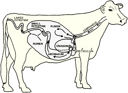
Anatomy of digestive tracts
Cattle have a digestive tract adapted to herbivore diet and to the rumen function, which gives a number of peculiarities. This tract allows the animals to ingest in short time considerable amounts of forage that are swallowed after a brief chewing, stored in a special compartment of the stomach, returned after a while again in the mouth, and again chewed and reswallowed. The digestive tract consists of the mouth, pharynx, esophagus, located in the prediafragmatic region; stomach compartments represented by the rumen, reticulum, omasum and actual stomach or abomasums located in the post-diaphragmatic region; small and large intestine, as well as the glands with digestive role (salivary, liver, gallbladder, pancreas). |

The oral cavity, pharynx and esophagus
The oral cavity, pharynx and esophagus are designed to ensure prehension, mastication and swallowing of food and water, respectively the rumination of the mericic bolus.
Teeth are characterized by the absence of upper incisors, which are replaced by a cartilaginous burlet. The dental formula for the adult cattle is the following: 0/4 incisors, 0/0 canines, 3/3 premolars and molars 3/3.
The pre-stomach
The pre-stomach occupies a large part of the abdominal cavity.
The rumen
The rumen is the most voluminous segment, representing about 3/4 of the volume of the entire digestive tract. The rumen mucosa is thick and durable, with many papillae that are involved in absorption, but it does not present digestive glands.
The reticulum
The reticulum is the smallest compartment, and inside it is the esophageal gutter, a semichannel that connects cardia with the orifice reticulum-omasum, being able to extend the esophagus up the the omasum. The mucosa of the reticulum is thick and it presents several creases that form polyhedral compartments, providing the appearance of a honeycomb.
Although they differ anatomically, the reticulum and the rumen form a functional unit. The reticulum also communicates with the esophagus through a cardial orifice, a slot located at the cranial extremily of the esophagus drain. By its topography, the cardial orifice allows the fall of heavier particles from the esophagus to the reticulum, which can boost the production of the traumatic reticulitis, especially through metal bodies. Moreover, the reticulum also communicates with the omasum through the reticulo-omasal orifice.
The esophagus
The esophagus drain is a trench long of 15-20 cm, located on the greater curvature of the reticulum between the cardial and the reticulo-omasal orifice. It is bordered by two edges made of longitudinal fibers of the mucosa, and the bottom of the trench is formed by crosslinking fibers of the reticular mucosa.
The omasum
The omasum exceeds the reticulum as volume, it has an ellipsoid shape, being craniocaudal flattened. It communicates to the left with the reticulum (reticulo-omasal orifice) and to the right and distal with the abomasums (omasoabomasal orifice). Between the two openings the omasal channel is placed, being located on the small curvature of the omasum and which connects the reticulum with the abomasums. The mucosa of the great curvature of the omasum is provided with numerous slides ranked in four size categories, structures that amplify the mucosal surface and that help in crushing the food intake.
Abomasum or the stomach
Abomasum or the stomach of ruminants itself, of reduced capacity (about 7% of the gastric complex) is located to the right and distal to the other compartments of the gastric complex. It shows a lower region, a body and a pyloric region. Its mucosa presents three different areas: the cardia glands region (reduced expansion, located near the omasoabomasal orifice), the area of the bottom glands and pyloric glands area. The pyloric orifice provides the communication between the abomasums and the duodenum, around this orifice existing a muscle ring with the role of a sphincter that separates through its contractions the pyloric region of the abomasums duodenum.
The small intestine
The small intestine is a long pipe of more than 30 m, strongly circumvolutionated, with constant diameter, but relatively small. The small intestine has three parts: duodenum (the initial part in U-shaped loop), jejunum (the largest portion, with numerous loops) and ileum (portion of the terminal, which communicates with the cecum through the ileocecolic orifice).
The intestinal mucosa forms folds (villi) that increase the absorption surface and the epithelium is composed of absorptive and endocrine cells. The thickness of the mucosa, particularly in the jejunum and ileum, there are lymphoid formations, lymph follicles and intestinal glands of serous type that secrete mucus or products having intense enzymatic activity. All these histological features contribute to the increase of exchange surface, favoring thus the absorption of nutrients.
The large intestine
The large intestine, having 8-11 m in length, extends from the ileum to the anus, with three parts: cecum, with a length of 0.6 to 1 m and the capacity of 5-6 liters; colon, the longest, floating, with three portions (ascending, transverse and descending colon); rectum, the terminal portion of the large intestine, characterized by the lack of haustrals folds and almost straight trajectory, which communicates with the outside through the anus.


